
Abstract
SMITracker was initiated by researchers in the Structural Biology and DNA Repair group at the Institute of Clinical Medicine, University of Oslo. The project aimed to develop an interactive computational platform for the management and analysis of DNA-scanning single-molecule data. A major practical bottleneck in single-molecule imaging is data analysis: experiments can generate millions of high-frequency image frames, but extracting reliable interaction trajectories and removing noise in a consistent and unbiased way is difficult and time-consuming. SMITracker addresses this bottleneck by converting preprocessed imaging data into structured R-compatible datasets, automating trajectory detection and noise exclusion, and producing diffusion-oriented outputs and visualizations that allow comparison across data from different proteins or experimental conditions. The tool is distributed both as an R package and as a Docker image [https://zenodo.org/records/18877064], making it usable for the R user community and beyond. For more details on SMITracker, please visit the recently published article in Computational and Structural Biotechnology Journal [https://spj.science.org/doi/10.34133/csbj.0014].
Background
Single-molecule experiments are important because they make it possible to observe molecular interactions directly, rather than only through ensemble averages. In the biological setting of this project, the target problem is how proteins interact dynamically with long, linear macromolecular substrates, especially DNA. This is directly relevant to DNA repair and genome maintenance research.
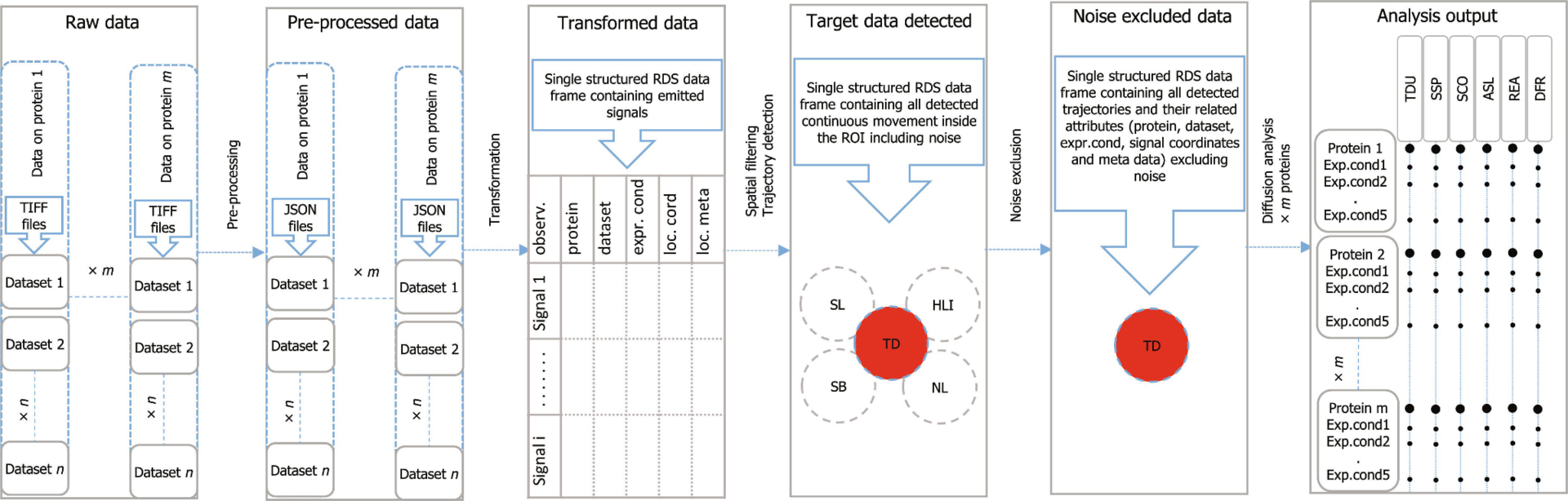
The computational challenge is that the imaging data are large, noisy, and sequential. A useful analysis system must therefore do more than detect bright spots in images. It has to connect localizations across time, determine which detections represent biologically meaningful molecular trajectories, support quality control, and return quantitative descriptors such as scanning speed, trajectory duration, and diffusion behavior. Such a complex task cannot be addressed with only an analysis script; it requires a deployable research platform for turning raw microscopy-derived data into curated, reproducible, and interpretable outputs. As shown in Figure 1, this process consists of multiple data-wrangling and analysis steps.
Methodology
The workflow begins with a preprocessing step for signal localization from recorded microscopy images. Signals in the raw images are first localized in Fiji using the ThunderSTORM single-molecule localization plugin. This preprocessing can be performed manually through the ThunderSTORM interface or automatically using a script provided with SMITracker. The result is then exported as JSON-formatted imaging data, which becomes the structured input for SMITracker.
Inside SMITracker, the computational pipeline is organized as a sequence of modules: data loading, spatial filtering, trajectory detection, visual inspection, noise exclusion, and analysis output. This modular design is important because each step transforms the dataset while preserving traceability between the raw localizations, filtered candidate signals, detected trajectories, manually reviewed subsets, and final analysis products.
The core computational contributions are the automatic detection of molecular trajectories and the systematic noise-exclusion model. SMITracker combines algorithmic event detection with standardized filtering to separate target molecular interactions from imaging artifacts or background signals. This is particularly important for high-throughput single-molecule imaging, where manual trajectory selection would be extremely time-consuming and potentially biased.
Although the main time-consuming tasks are automated, the platform also includes human-in-the-loop validation throughout the processing workflow. This helps ensure analysis integrity while still benefiting from efficient and automated pipelines. Figure 2 shows an example of a processed dataset in which the target data are visualized together with different types of noise. The user can visualize the results of the trajectory detection and noise exclusion modules separately, providing both a bird’s-eye view of the process and the possibility of examining the operations and results in greater detail.

SMITracker goes beyond trajectory detection. In the final module, it can extract various biologically relevant quantitative variables from the detected data, including scanning speed, binding lifetime, scanning coverage, accumulated scanning length, redundancy versus efficiency, and diffusion rate. In addition, this module allows users to accumulate data from different proteins and different analysis rounds, enabling side-by-side comparison of proteins across these variables. Finally, the availability of SMITracker as both an R package and a Docker image provides flexibility for both R and non-R users.