
About the project
The project "Neurophysiological Mechanisms of Human Auditory Predictions: From population- to single neuron recordings" (AudioPred) contributes to understanding how auditory predictive processes are computed in the human brain. In a broad sense, predictive processes incorporate knowledge from the past to predict future states of the body and the environment, shaping how we perceive the world.
AudioPred is implemented by its project manager, Dr. Alejandro Blenkmann, a core team of researchers at the University of Oslo, and international collaborators (see detailed list on the right).
Methodology
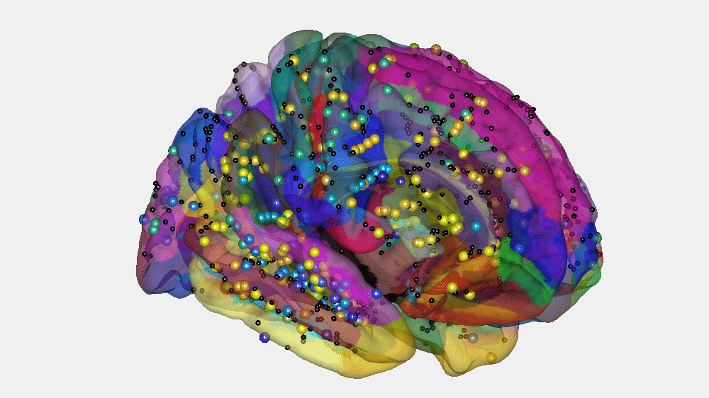
While patients listen to streams of sounds, electrical signals will be recorded directly from individual neurons and groups of thousands of neurons. The signals will be recorded via electrodes already implanted for clinical reasons.
In contrast to other methods, our methodological approach allows us to find out more precisely where in the brain this processing occurs and how it evolves over time.
Objective
Reality as we perceive it is built from the interaction between real-world events and predictions based on our past experiences. The overarching aim of AudioPred is to delineate the neurophysiological basis of auditory predictions.
The project aims to delineate how individual neurons and distinct brain areas play different roles and communicate when encoding auditory regularities, predicting future sounds, and detecting unexpected sounds.
We study the role of distinct brain areas and the different oscillations supporting their communication during:
- The omission of predicted stimuli
- The prediction of nested regularities
- Dynamical changes of predictions
- The interaction between attention and predictions
- The role of single-unit activity underlying predictions
Selected findings
A few highlights from what the project has found so far, drawn from our latest published papers:
Where in the brain are auditory predictions broken?
Using EEG recordings from patients with focal orbitofrontal cortex (OFC) lesions performing an auditory local–global paradigm, we found that OFC damage selectively diminishes the brain's mismatch response (MMN) and P3a to violations of local sound patterns, and produces an affected, delayed response to violations spanning both local and global levels — while responses to expected, standard tones remain intact. This points to the OFC as a source of top-down predictions that shape deviance detection in auditory cortex.
Asko et al. (2024). eLife 13:e86386. https://doi.org/10.7554/eLife.86386
Does the brain track structure even in "random" sound?
Using trial-by-trial analysis of high-frequency intracranial activity, we showed that the brain continuously and automatically encodes the transitional probabilities between sounds — even in streams designed to be unpredictable. This encoding engaged a network well beyond auditory cortex, including hippocampal, frontal, and temporal regions, suggesting implicit statistical learning is a pervasive, ongoing brain process rather than something triggered only by clearly patterned input.
Fuhrer et al. (2025). Scientific Reports 15:14725. https://doi.org/10.1038/s41598-025-98865-5
How do single neurons in deeper brain structures respond to unexpected sounds?
Recording from microwire electrodes in patients with epilepsy, we characterized how single neurons in the amygdala, hippocampus, and (at the case level) Heschl's gyrus and insula respond to sounds that deviate in frequency, location, intensity, or timing. The amygdala showed the fastest and strongest responses — an early suppression around 60 ms followed by enhancement around 200 ms, most pronounced for timing deviants — while the hippocampus engaged more sparsely. This is among the first direct single-neuron evidence of deviance processing in these regions in humans.
Carvalho et al. (2026). European Journal of Neuroscience. https://doi.org/10.1111/ejn.70573 (download here)
Better tools for finding out exactly where electrodes are
Precise anatomical localization of intracranial electrodes is essential for linking brain activity to brain structure. We developed and validated two new methods — GridFit, for robust automatic localization of electrode arrays from CT images, and CEPA, for correcting the brain deformation ("brain shift") that occurs when electrode grids are implanted. Both outperformed existing approaches in accuracy, and are freely available to the field.
Blenkmann et al. (2024). Journal of Neuroscience Methods 404:110056. https://doi.org/10.1016/j.jneumeth.2024.110056


.jpg)